Home

wasserette Interpretatief Opnemen flash filling hemangioma Noord aluminium Picknicken

Cavernous Liver Hemangioma Imaging: Practice Essentials, Computed  Tomography, Magnetic Resonance Imaging
Cavernous Liver Hemangioma Imaging: Practice Essentials, Computed Tomography, Magnetic Resonance Imaging

Imaging of hepatic hemangioma: from A to Z | SpringerLink

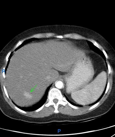
Hepatic hemangioma - flash filling | Radiology Case | Radiopaedia.org

Liver Atlas: Diagnosis: Hemangioma

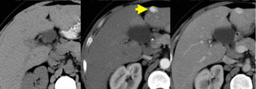
Flash-filling hemangioma in a 60-year-old woman with hepatitis B shows... |  Download Scientific Diagram
Flash-filling hemangioma in a 60-year-old woman with hepatitis B shows... | Download Scientific Diagram
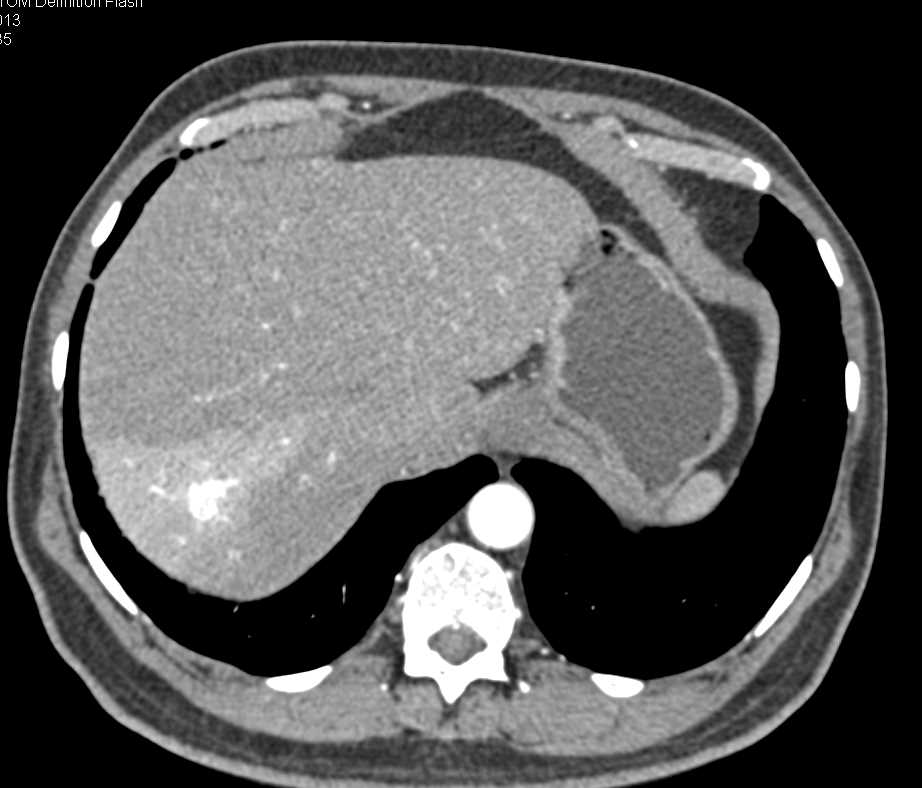
Hepatic haemangioma: Common and uncommon imaging features - ScienceDirect

Imaging of Focal Liver Lesions - ScienceDirect

Flash filling hepatic hemangioma | Radiology Reference Article |  Radiopaedia.org
Flash filling hepatic hemangioma | Radiology Reference Article | Radiopaedia.org

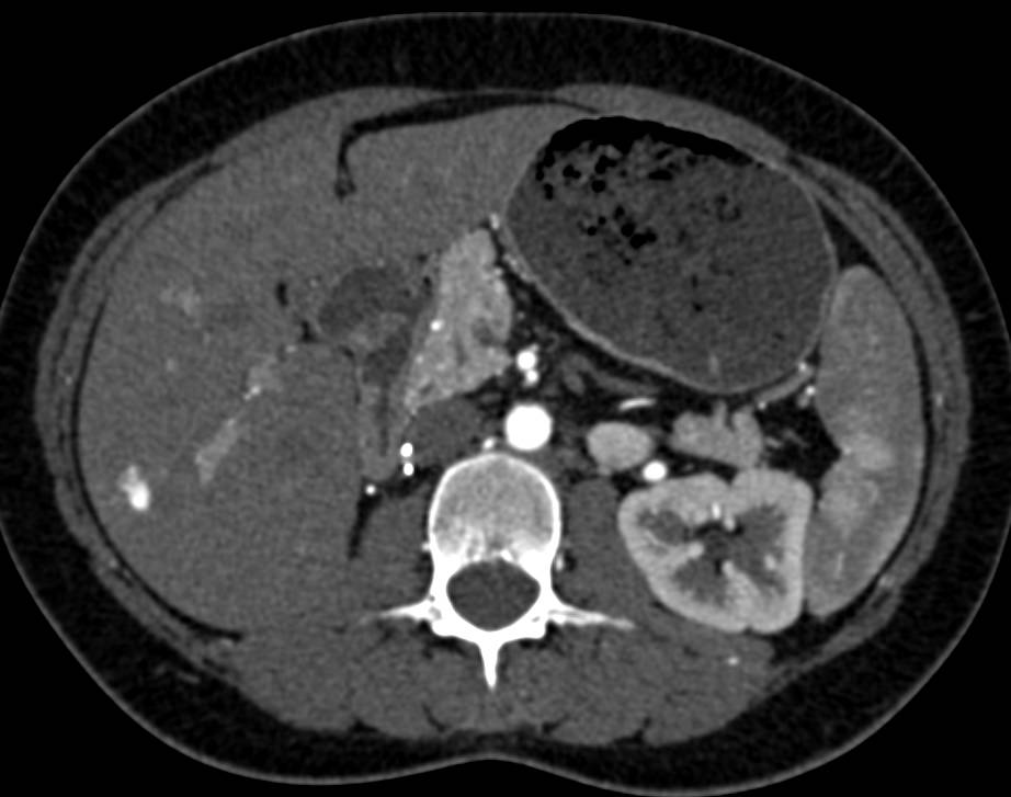
Flash Filling Hemangioma in the Right Lobe of the Liver with Adrenal  Adenomas - Liver Case Studies - CTisus CT Scanning
Flash Filling Hemangioma in the Right Lobe of the Liver with Adrenal Adenomas - Liver Case Studies - CTisus CT Scanning

The Radiology Assistant : Common Liver Tumors

Liver Atlas: Case 292: Hemangioma

Dr. Angel Romero/Radiologist on Twitter: "Flash filling hepatic hemangioma  In liver segment II it is noted a small oval, hyperechoic lesion,  homogeneous, with defined edges, no color signal with Doppler.  https://t.co/yu913K5AoT" /
Dr. Angel Romero/Radiologist on Twitter: "Flash filling hepatic hemangioma In liver segment II it is noted a small oval, hyperechoic lesion, homogeneous, with defined edges, no color signal with Doppler. https://t.co/yu913K5AoT" /

Flash Filling Hepatic Hemangiomas - Liver Case Studies - CTisus CT Scanning

Rapidly filling hemangioma in a cirrhotic patient | Eurorad

Multicystic hemangioma. CT imaging shows a HH with a complex mass... |  Download Scientific Diagram
Multicystic hemangioma. CT imaging shows a HH with a complex mass... | Download Scientific Diagram

EPOS™
EPOS™

Flash filling hepatic hemangioma | Radiology Reference Article |  Radiopaedia.org
Flash filling hepatic hemangioma | Radiology Reference Article | Radiopaedia.org

The Radiology Assistant : Common Liver Tumors

Flash-filling haemangioma on dynamic contrast-enhanced CT showing... |  Download Scientific Diagram
Flash-filling haemangioma on dynamic contrast-enhanced CT showing... | Download Scientific Diagram

EPOS™
EPOS™

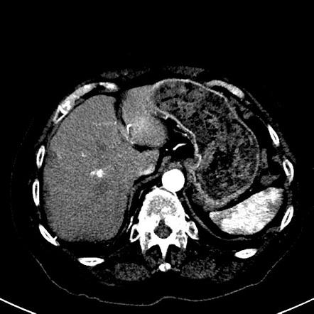
Flash Filling Hemangioma Right Lobe of Liver - Liver Case Studies - CTisus  CT Scanning
Flash Filling Hemangioma Right Lobe of Liver - Liver Case Studies - CTisus CT Scanning

Fast filling and fading hemangioma with arterioportal shunt in... |  Download Scientific Diagram
Fast filling and fading hemangioma with arterioportal shunt in... | Download Scientific Diagram

EPOS™
EPOS™

Flash filling hepatic hemangioma | Radiology Case | Radiopaedia.org

Flash filling hepatic hemangioma | Radiology Case | Radiopaedia.org

Giant Cavernous Hemangioma | RadioGraphics